Search This Blog

Follow me


Monday, August 14, 2023


 **Narrative Article on Coccyx Physiotherapy Treatment: A Comprehensive Guide**


In the world of physiotherapy, there's a crucial area that often goes unnoticed but can cause significant discomfort and hinder daily activities if not properly addressed: the coccyx, commonly known as the tailbone. At our specialized clinic, we understand the importance of coccyx health and the impact it has on overall well-being. In this comprehensive guide, we'll delve into the intricacies of coccyx issues and how effective physiotherapy treatment can alleviate pain, improve mobility, and enhance your quality of life.


## Understanding the Coccyx


The coccyx is a small triangular bone located at the base of the spine. It consists of three to five fused vertebrae and plays a crucial role in supporting the body while sitting and providing attachment points for various muscles, tendons, and ligaments. Due to its location and function, the coccyx is susceptible to injuries, inflammation, and chronic pain.


## **Common Coccyx Issues**


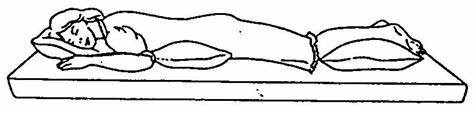
Several factors can contribute to coccyx-related problems, including trauma from falls, prolonged sitting on hard surfaces, childbirth, or even chronic conditions such as coccydynia (persistent coccyx pain). Individuals experiencing coccyx pain often find it challenging to sit, stand, or engage in physical activities, severely impacting their daily routines and overall comfort.


## **The Role of Physiotherapy**


Physiotherapy is a non-invasive, holistic approach that can significantly alleviate coccyx pain and address related issues. Our experienced physiotherapists employ a personalized approach to assess each patient's condition, create tailored treatment plans, and monitor progress throughout the recovery journey.


### **1. Assessment and Diagnosis**


Our first step is a comprehensive assessment to understand the root cause of the coccyx issue. We consider factors such as medical history, lifestyle, previous injuries, and the specific nature of the pain. This thorough evaluation allows us to pinpoint the underlying problem accurately.


### **2. Tailored Treatment Plans**


Once we've identified the cause of the discomfort, we design a customized treatment plan that may include a combination of manual therapies, gentle exercises, postural adjustments, and ergonomic advice. Our focus is not just on relieving immediate pain but on addressing the root issue to prevent future occurrences.


### **3. Gentle Exercises and Rehabilitation**


Engaging in appropriate exercises is vital for the recovery process. Our physiotherapists guide patients through gentle exercises that strengthen the surrounding muscles, improve flexibility, and enhance overall spinal health. These exercises are crucial for the long-term well-being of the coccyx and the entire spine.


### **4. Pain Management Techniques**


We understand that managing pain is a significant concern for our patients. Our team employs various pain relief techniques, including heat and cold therapy, gentle massage, and relaxation techniques, to alleviate discomfort and promote healing.


### **5. Lifestyle Modifications**


In addition to the direct treatment of the coccyx, we provide valuable guidance on making necessary lifestyle modifications. This includes recommendations for proper sitting posture, using supportive cushions, and incorporating regular movement breaks, especially if you have a sedentary job.


## **Your Journey to Coccyx Wellness**


At our clinic, we're committed to empowering our patients with the knowledge and tools they need to regain coccyx health and overall well-being. Our approach goes beyond short-term relief; we aim for long-lasting results that enable you to live your life to the fullest, free from the constraints of coccyx pain.


If you're ready to embark on a journey towards coccyx wellness and enjoy a life free from discomfort, don't hesitate to reach out to our dedicated team of physiotherapists. We're here to support you every step of the way, helping you regain mobility, comfort, and confidence.


## **Conclusion**


In this comprehensive guide, we've explored the significance of coccyx health, common issues, and the pivotal role of physiotherapy in addressing coccyx-related discomfort. Our personalized approach, tailored treatment plans, and commitment to long-term well-being set us apart as leaders in coccyx physiotherapy.


Wednesday, June 9, 2021

Movement


Movement perform and controlled by the voulantary action of muscle, working in oppsition to an external force.

CLASSIFICATION:- 


- Free exercise

        - Assisted exercise

       - assisted resisted Exercise

        - Resisted exercise


free exercise. The working muscles are subjected only to the forces of gravity acting upon the part moved or stabilised.

Assisted exercise:

when muscle strength or coordination is in-adequate to perform a movement an external force is applied to compensate for the deficiency.

ASSISTED - RESISTED EXERCISE:-

Muscle may be strong enough to work against resistance in part of the range and not in other. this type of exercise ensures that the external forces applied are adapted are adapted in every part of the abilities of the muscles.


 Resisted exercises: The force of resistance offered to the action of the working muscles are artificially and systematically increased to develop the power and endurance of the muscle.

Sunday, May 23, 2021

WATCH OUT Mucormycosis IN COVID -19 PATIENT

 HIGH suspecious:

- Diabetic melliyus 

- Treatment for covid -19 with corticosteriods.


Treatment with immuno moddulators(Tocilizumb,itolizumb).


- Treatment with Mechanical ventilation.


- long standing oxygen therapy.


- EARLY SIGN ANS SYMPTOMS:


- facial swelling 

- facial skin discoloration

- ptosis

- proptosis's

- ristricted eye movement 

- palate discoloration

- palatal Necrosis

- Brownish dischrage from nose


- facial pain 

- sinus headache

- stuffy nose

- bloody nasal discharge

- blurring of vision 

- dental pain 

- loosening of teeth


EARLY DETECTION AND EARLY INTERVENTION:

- CBC

- blood sugar

- renal function tests (s,creatnine BUN)

- Deep nasal swab-KOH stai<b></b>ning

- nasal endoscopy biopsy

 - MRI-for extent invoved

- CT scan-PNS To determine extent of involvement


BEST OUTCOME:

REDUCE morbidity and mortality

- medical managment

- strict blood sugar

- antifungals

- amphotericin B1.5mg/kg/dayfor 4-6weeks

- posaconzazole loading dose 300mg BD,300mgOD FOR 4-6 weeks

- Early surgical Debridement


maxillofacial surgeon

ENT Reference

Opthamological reference

Neurological reference.


Sunday, May 16, 2021

Managing covid-19 at home Have been Advice for home quarantine due to mild symptoms.


As peradvisery guidance of govt of India ministry of health patients with mild covid infection can be managed at home only. with symptomatic control medication and with proper care we know that covid -19, 70-80% infection would be mild you will get better within few days to few weeks your doctor will give you the best treatment with taking care of symptom and help you get better fast

Here we talking about some aspect which we will enhance speedy recovery:

Take proper rest

To maintain the daily routine

Get up on time, Shower once a day, change your clothes daily and maintain proper hygiene, maintain a level of activity, move above 10-15 minute 2-3 times in your room every day, this will keep you refresh, it will keep the circulation leg your going and prevent the chances of deep vein thrombosis which can happen because of prolonging resting.

Diet should be easy to digest but should have all the required nutrients (cereal, protein, fruits which rich in vitamin c citric fruit and sessional fruit will very help full for your recovery, the importance of liquids and water cannot be over emphasizes have about 3-4 liters of liquid, warm water, tea coffee, milk, etc, also you can have turmeric, honey, ginger,giloy this thing in your diet as a supplement to your nutritional food requirement as well as to boost your immunity. because covid 19  infection you are through main scratchy irritated and sometimes even pain is important to do gargle at least 3 times a day after eating your food to reduce pain, irritation, viral load in throat secretions.

During covid 19 infection the main organ which takes the brants is your lungs to keep your lung in good shape do this thing:


1-do steam at least three times a day.


2-try to lying down on your stomach, pillow placement is very important when you are doing this don’t press your stomach because between the chest and hip bone our diaphragmatic muscle is lying don’t press your stomach diaphragmatic movement is very important. place one pillow beneath the upper chest and another on beneath the hip pone and you can move your hand as comfortable you are. it will improve and open up your lungs and the back of the chest where most of the part of the lung is lying. so it will improve oxygenation so while resting and be careful don’t do it immediately after eating food it is also contraindicated for obese( who have a fatty stomach, it can disturb diaphragm function ), pregnancy, spondylolisthesis, back surgery, thigh bone surgery you should consult with you doctor before doing this.


3.some deep breathing exercise.

That can help with a spirometer or you can take the help of normal birthday balloons, blowing to the balloon for good 3 breaths and holding some time, inhale and take out the breath very gradually, it will keep your lungs in good shape and it will improve the oxygenation. And can also do deep breathing or breath-holding exercise if you are doing the first time you may hold 5 sec and increase your holding 2-3 sec daily with your family member and encourage to each other to increase your breath holding time it will maintain your lung capacity and shape.


4-Mental health is equally important in covid 19infection for speedy recovery purpose don’t panic that is worst you can do stay very positive stay away from negativity don’t watch too much of negative news on television rather invest your time in resting, reading good books, listen to good and piece full music, and if time permits connect with your friends through phone and video chat keep positivity around you don’t panic at all this will help very much in your speedy recovery.

Saturday, November 28, 2020

Manual muscle test (MMT)

Manual muscle test (MMT)
1. MANUAL MUSCLE TEST (MMT)
INTRODUCTION:  Manual muscle testing is employed to work out the extent and degree of muscular weakness resulting from disease, injury or disuse. The records obtained from these tests provide a base for planning therapeutic procedures and periodic re-testing. Muscle testing is a crucial tool for all members of health team handling physical residuals of disability.
Muscular strength: The maximal amount of tension or force that a muscle or muscle group can voluntarily exert during a maximal effort; when sort of contraction , limb velocity and joint angle are specified.  Muscular endurance: the power of a muscle or a muscle group to perform repeated contractions against resistance or maintain an isometric contraction for a period of your time . 
Muscle power:  Power is defined because the generate the maximum amount force as fast as possible.  Power does require strength and speed to develop force quickly.  POWER = strength speed.
TYPES OF MUSCLE WORK: 1. Isometric contraction: Tension is developed within the muscle but no movement occurs; the origin and insertion of the muscle don't change their positions and hence, the muscle length doesn't change. 2. Isotonic contraction: The muscle develops constant tension against a load or resistance. There are two types: a) Concentric contraction: Tension is developed within the muscle and therefore the origin and insertion of the muscle move closer together; therefore the muscle shortens. b) Eccentric contraction: Tension is developed within the muscle and therefore the origin and insertion of the muscle move further a part; therefore the muscle lengthens.
RANGE OF MUSCLE WORK: the complete home in which a muscle work refers to the muscle, changing from an edge of full stretch and contracting to an edge of maximal shortening. the complete range is split into three parts: 1. Outer range: From an edge where the muscle is fully stretched to an edge halfway through the complete range of motion. 2. Inner range: From an edge halfway through the complete range of motion to an edge where the muscle is fully shortened. 3. Middle range: The portion of the complete range between the mid-point of the outer range and therefore the midpoint of the inner range.
GROUP OF MUSCLE ACTION: 1. first cause or agonist: A muscle or muscle group that creates the main contribution to movement at the joint. 2. Antagonist: A muscle or a muscle group that has an opposite action to the prime movers. The antagonist relaxes because the agonist moves the part through a variety of motion. 3. Synergist: A muscle that contracts and works along side the agonist to supply the specified movement.
There are three sorts of synergists: a) Neutralizing or counter-acting synergist b) Conjoint synergist c) Stabilizing or fixating synergist a) Neutralizing or counter-acting synergists: Muscles contract to stop any unwanted movement produced by the first cause . for instance , when the long finger flexors contract to supply finger flexion, the wrist extensors contract to stop wrist flexion from occurring.
b) Conjoint synergists: Two or more muscles work together to supply the specified movement. for instance , wrist extension is produced by contraction of extensor carpi radialis longus, carpi radialis brevis and extensor carpi ulnaris muscles. If the extensor carpi radialis longus or brevis contracts alone, the wrist extends and radially deviates, while if the extensor carpi ulnaris contracts alone, the wrist extends and ulnarly deviates. When the muscles contract as a gaggle , the deviation action is cancelled, and therefore the common action occurs.
c) Stabilizing or fixating synergists: These muscles prevent or control the movement at joints proximal to the moving joint to supply a hard and fast or stable base, from which the distal moving segment can effectively work. for instance , if the elbow flexors contract to lift an object off a table anterior to the body, the muscles of the scapula and gleno-humeral (shoulder) joint must contract to either allow slow controlled movement or no movement to occur at the scapula and gleno-humeral joint to supply the elbow flexors with a hard and fast origin from which to tug . If the scapular muscles don't contract, the thing can't be lifted because the elbow flexors will act to tug the pectoral arch downward.
  1. 2) DEFINITION OF MMT:  Manual muscle test (MMT) could even be a procedure for the evaluation of strength of individual muscle or muscles group, based upon the effective performance of a movement in regard to the forces of gravity or Manual Resistance through the available Range of motion (ROM).  MMT is that the foremost significant a neighborhood of motor assessment performed in checkup .
  2. BASIC COMPONENTS OF MOTOR EXAMINATION: i. Nutrition or Bulk of muscle ii. Tone iii. Reflexes iv. Range of motion (ROM) v. Manual muscle test (MMT) vi. Functional Assessment *Important of the Sequence
  3. Individual versus group muscle test:  Muscles with a typical action or actions could even be tested as a gaggle or each muscle could even be tested individually. for instance , flexor carpi ulnaris and flexor carpi radialis muscles could even be tested together as a gaggle in wrist flexion. Flexor carpi ulnaris could even be tested more specifically within the action of wrist flexion with ulnar deviation. On the other hand, Flexor carpi radialis longus and brevis could even be tested more specifically within the action of wrist flexion with radial deviation.
  4. 3) Purposes and uses of MMT:  CLINICAL USES : i. The severity of problem are often understand. (It is diagnostic Tool) ii. we will planning our treatment goals. iii. Determine the extend & degree of muscular weakness resulting from disease, injury. iv. Correlating muscle picture with in level innervations (myotoms) . v. MMT may be a crucial tool for all the members of the Rehabilitation team. vi. Prevents deformities by locating problem areas. vii. Help and Evaluate effectiveness of treatment to the therapist.
  5. WHY MMT IS PERFORMED? : to urge some answers such as:- i. may be a particular muscle is normal? ii. Is it weak? (How much weak) iii. Is it strong enough? (How much strong) iv. Is it weak on both the side (bilateral symmetrical)? v. Is it weak only on one side (Unilateral)? vi. Is proximal muscles are weaker than the proximal one? vii. Is there any particular pattern of muscle weakness?
  6. GRADES OF MMT: i. MRC Scale ii. OXFORD Scale iii. KENDALL Scale iv. And Others .
  7. MEDICAL RESEARCH COUNCIL (MRC) SCALE:
  8. OXFORD SCALE :
  9. KENDALL SCALE:
  10. + or – GRADES :
  11. 5) PRINCIPLES OF MMT: 1) Position 2) Stabilization 3) Demonstration 4) Application of Grades 5) Application of Resistance 6) Checking normal strength 7) Objectivity 8) Documentation 1) POSITION : PATIENT POSITION:  Patient is positioned Eliminated or Against gravity. (Patient depend on testing on muscle or muscles group).  Do not change patient position repeatedly.  The patient should be as free as possible from discomfort or pain for the duration of every test. It may be necessary to permit some patients to maneuver or be positioned differently between tests.  Patient position should be carefully organized so that position changes in a test sequence are minimized. The patient' s position must permit adequate stabilization of the part or parts being tested by virtue of weight or with help provided by the examiner.
  12. JOINT POSITION:  The joint position is also changed depend upon their performance.  Distal part of the joint is moved.  Place the joint in Antigravity position- Grade 3  Place the joint in Horizontal position – Grade 4
  13. 2) STABILIZATION :  Patient could stabilizes our self during performed Antigravity position.  The hand placement of the therapist is important. HAND PLACEMENT: I. PROXIMAL HAND – At Origin of muscle & proximal joint giving stabilization. II. DISTAL HAND – Distally offering resistance or Assistance depend on performance. 3) DEMONSTRATION:  Demonstrate the desired movement.  Therapist demonstrate the appliance of movement or performance to the patient. 4) APPLICATIONS OF GRADES:  Always start with GRADE 3 (If you start to examine the muscle power, first you should test the grade 3).  Isolation of muscle could be tested.
  14. d) External forces: They may be applied manually by the therapist or mechanically by devices such as belts and sandbags.  e) Substitution and trick movements: When muscles are weak or paralyzed, other muscles may take over or gravity may be used to perform movements normally carried out by the weak muscles.
  15. 5) APPLICATIONS OF RESISTANCE:  Resistance is applied slowly & gradually.  Increasing or decreasing manual resistance.  Increasing length of weight arm.  Apply presence opposite to the road of pull (Grade 4,5)  Apply force distally.  It varies between the persons.  Use long lever to applied resistance whenever it possible.
  16. . 6) CHECKING NORMAL STRENGTH:  Therapist to check the strength of the muscle normal side first. 7) OBJECTIVITY:  Therapist ability to palpate and observe the tendon or muscle response in very weak muscles.
  17. . 8) DOCUMENTATION:  Examiners complete testing documentation or Record first.  this may help for next step of treatment applications.  And help for checking improvement of treatment.
  18. 7) INDICATIONS OF MMT: 1)Lower efferent neuron (LMN) Disease. 2) Some other Neurological (Neuromuscular )disease. Such as,  MS  Muscular distrophy  Amyotropic Lateral Sclerosis  myasthenia .  Guillian - barre syndrome (GBS), etc.... 3) Some Musculoskeletal disorders.
  19. 8) CONTRAINDICATIONS OF MMT: 1) Cerebral Palsy 2) Cardio vascular disease / Brain injury 3) Dislocated/ unhealed fracture 4) Myositis ossifications 5) Parkinson’s disease 6) Pain 7) Inflammation /(inflammatory disease in muscles and or joints) 8) Severe cardiac & respiratory disease .
  20. . Cont. 9) Subluxation joint 10) Hemophelia 11) Osteoporosis
  21. . 9) PRECAUTION: 1) Considered contraindications 2) don't harm (Be gentle) 3) Respect pain 4) Examiner know the available ROM. 5) Follow the principles of procedure 6) lookout of patient comfort 7) Record accurately. 8) Extra care taken to giving Resisted Exercise.
  22. Cont. 9) Abdomen surgery or hernia 10) Newly united fracture 11) Bony ankylosis 12) Hematoma 13) If patients take muscle relaxers and or pain medications 14) Prolonged immobilization
  23. Extra care must be taken where Resisted movements might aggravate the condition:  Patients with history in danger of getting cardiovascular problems.  Abdominal surgery or herniation of wall to avoid stress on the wall .  Fatigue exacerbate the patients condition.  Patient with extreme debility, for instance ,  Malnutrition  Malignancy  And Severe COPD.
  24. 10) LIMITATION OF MMT: 1) UMN LESIONS : Spastic muscle have poor control from higher centers thus its better to go for voluntary control assessment rather than MMT. 2)RESTICTED ROM DUE TO TCD’S (Transcranial Doppler) : TCD’s can give faulty interpretation about MMT, thus in case always mention about the MMT within available range along with Grade. 3) PRESENCE OF PAIN & SWELLING: pain and swelling increases the intra articular tension causing irritation of joint and can affect the MMT result, thus in case always mention about presence of pain along with Grade.
  25. 4) TYPES OF CONTRACTION : MMT gives idea about Quality of concentric contraction only. (Not Eccentric which is more functional). 5) UNDERSTANDING OF COMMANDS:  Paediatric Age group < 5 years  IQ  Higher functions. 6) STRENGTH Vs ENDURANCE: MMT give knowledge about only the strength and not endurance.
  26. 7) Subjectivity (patient) HOOVERS sign 8)And following methods also Limit the MMT ;  Showing the Co-ordination  Showing pictures of gross / patient contraction  Showing the ability of client to use muscle power  Showing the how much joint ROM the individual is working through.
  27. 11) PROCEDURE: 1) Explanation & Instruction 2) Assessment of normal muscle strength 3) Properly positioned the patient 4) Stabilization 5) Substitution movements & Trick movements 6) Conventional methods 7) Alternating techniques.
  28. PREPARATIONS:
  29. The plinth or mat table for testing must be firm to assist stabilize the part being tested. The ideal may be a pave , minimally padded or not padded in the least . The pave won't allow the trunk or limbs to "sink in. " Friction of the surface material should be kept to a minimum. When the patient is fairly mobile a plinth is ok , but its width shouldn't be so narrow that the patient is scared of falling or sliding off. When the patient is severely paretic, a mat table is that the more practical choice. The height of the table should be adjustable to allow the examiner to use proper leverage and body mechanics.
  30. Materials needed include the following: • Muscle test documentation forms • Pen, pencil, or computer terminal • Pillows, towels, pads, and wedges for positioning • Sheets or other draping linen • Goniometer • Interpreter (if needed) • Assistance for turning, moving, or stabilizing the patient • Emergency call system (if no assistant is available) • Reference material
  31. 1) EXPLANATION & INSTRUCTION: The therapist demonstrate and or explains briefly the movement to be performed and or passively moves the patient’s limb through the test movement. 2) ASSESSMENT OF NORMAL MUSCLE STRENGHT: Always assess and record the strength of the unaffected side limb to determined the patient’s normal strength.
  32. 3) PROPERLY POSITINED THE PATIENT: The patient is positioned to isolate the muscle (or) muscles group to be tested in either gravity eliminated or Against gravity positioned.
  33. 3) STABILIZATION: I. PROXIMAL HAND – At Origin of muscle & proximal joint giving stabilization. II. DISTAL HAND – Distally offering resistance or Assistance depend on performance.  The plinth or mat table for testing must be firm to assist stabilize the part being tested.
  34. The site of attachment of the muscle origin should be stabilized, therefore the muscle features a fixed point from which to tug . Substitutions and trick movements are avoided by making use of the subsequent methods: a) The patient's normal muscles: for instance , the patient holds the sting of the plinth when hip flexion is tested and uses the scapular muscles when gleno- humeral flexion is performed. b) The patient's body weight: it's wont to help fix the proximal parts (shoulder or pelvic girdles) during movement of the distal ones. c) The patient’s position: for instance , when assessing hip abduction strength in side lying, the patient holds the non-tested limb in hip and knee flexion so as to tilt the pelvis posteriorly and to repair the pelvis and lumbar spine.
  35. 4) CONVENTIONAL METHODS:  Manual grading of muscle strength is predicated on three factors: * Evidence of contraction: No palpable or observable contraction (grade 0) or a palpable or observable contraction with no joint motion (grade 1). * Gravity as a resistance: the power to maneuver the part through the complete available range of motion with gravity eliminated (grade 2) or against gravity (grade 3). * Amount of manual resistance: the power to maneuver the part through the complete available range of motion against gravity and against moderate manual resistance (grade 4) or maximal manual resistance (grade 5). * Adding (+) or (-) to the entire grades: this is often needed to denote variation within the range of motion. Movement through but half the available range of motion is denoted by a “+” (outer range), while movement through greater than half the available range of motion is denoted by “-“ (inner range).
  36. CONVENTIONAL GRADING:
  37. Numerals Letters Description Against gravity tests: The patient is in a position to maneuver through: 5 N (normal) the complete available ROM against gravity and against maximal manual resistance, with hold at the top of the ROM (for about 3 seconds). 4 G (good) the complete available ROM against gravity and against moderate manual resistance. 4- G - (good -) Greater than one half the available ROM against gravity and against moderate manual resistance. 3+ F + (fair +) but one half the available ROM against gravity and against minimal manual resistance. 3 F (fair) the complete available ROM against gravity. 3- F - (fair -) Greater than one half the available ROM against gravity. 2+ P + (poor +) but one half the available ROM against gravity. Gravity eliminated tests: The patient is in a position to actively move through: 2 P (poor) the complete available ROM with gravity eliminated. 2- P - (poor -) Greater than one half the available ROM with gravity eliminated. 1+ T + (trace +) but one half the available ROM with gravity eliminated. 1 T (trace) None of the available ROM with gravity eliminated and there's palpable or observable flicker contraction. 0 0 (zero) None of the available ROM with gravity eliminated and there's no palpable or observable muscle
  38. SCREENING TEST:  A audition may be a method wont to control muscle strength assessment, avoid unnecessary testing and avoid fatiguing and / or discouraging the patient. The therapist may screen the patient through the knowledge gained from: 1. The previous assessment of the patient's active range of motion. 2. Reading the patient's chart or previous muscle test result. 3. Observing the patient while performing functional activities. for instance , shaking the patients hand may indicate the strength of grasp (finger flexors). 4. All muscle testing procedures must begin at a specific grade; this is often usually grade “fair”. The patient is instructed to actively move the part through full range of motion against gravity. Based upon the results of this first test, the muscle test is either stopped or proceeds.
  39. FACTORS AFFECTING STRENGTH: 1). Age: A decrease in strength occurs with increasing age thanks to deterioration in muscle mass. Muscle fibers decrease in size and number; there's a rise in animal tissue and fat and therefore the respiratory capacity of the muscle decreases. Strength apparently increases for the primary 20 years of life, remains at this level for five or 10 years then gradually decreases throughout the remainder of life. The changes in muscular strength by aging are different for various groups of muscles. The progressive decrease in strength is clearer within the forearm flexors and muscles that raise the body (anti-gravity muscles).
  40. 2). Sex: Males are generally stronger than females. The strength of males increases rapidly from 2 to 19 years aged at a rate almost like weight and more slowly and frequently up to 30 years. then , it declines at an increased rate to the age of 60 years. The strength of females is found to extend at a more uniform rate from 9 to 19 years and more slowly to 30 years, after which it falls off during a manner almost like males. it's been found that ladies are more 28 to 30% weaker than men at 40 to 45 years aged .
  41. . 3). sort of muscle contraction: More tension are often developed during an eccentric contraction than during an isometric contraction. The concentric contraction has the littlest tension capability. 4). Muscle size: The larger the cross-sectional area of a muscle, the greater the strength of this muscle are going to be . When testing a muscle that's small, the therapist would expect less tension to be developed instead of if testing an outsized , thick muscle.
  42. 5). Speed of muscle contraction: When a muscle contracts concentrically, the force of contraction decreases because the speed of contraction increases. The patient is instructed to perform each movement during muscle test at a moderate pace. 6). Previous training effect: Strength performance depends abreast of the power of the systema nervosum to activate the muscle mass. Strength may increase together becomes conversant in the test situation. The therapist must instruct the patient well, giving him a chance to maneuver or be passively moved through the test movement a minimum of once before muscle strength is assessed.
  43. 7). Joint position: It depends on the angle of muscle pull and therefore the length-tension relationship. the strain developed within a muscle depends upon the initial length of the muscle. no matter the sort of contraction , a muscle contracts with more force when it's stretched than when it's shortened. the best amount of tension is developed when the muscle is stretched to the best length possible within the body (if the muscle is fully outer range). 8). Fatigue: because the patient fatigues, muscle strength decreases. The therapist determines the strength of muscle using as few repetitions as possible to avoid fatigue.
  44. The patient's level of motivation, level of pain, body type, occupation and dominance are other factors which will affect strength.
  45. BREAK TEST:  Resistance applied at the top of tested range is termed as BREAK TEST.  For one joint muscle resistance is applied at End of ROM.  for 2 joint muscle resistance is applied at Mid Range.  The isometric hold (break test) shows the muscle to possess a better grade than the make test.  MAKE TEST: Resistance is applied throughout the test is called MAKE TEST.
  46. INDICATIONS OF BREAK TEST: 1. When movement is contraindicated 2. When there is pain in movement 3. When we need to assess the standard of strength and not the quantity?.
  47. INTRUMENTATION:  Instrument chosen to assess muscle strength depends on the degree of accuracy required within the measurement.
  48. HAND HELD DYNAMOMETER: This Device operate on principle of compression. Application of external force to the dynamometer compress a steel spring and moves a pointer.
  49. PINCH GAUGE: pinch may be a strength measurement using pinch dynamometer.
  50. CABLE TENSIOMETER: Force during knee extension increased force on cable depresses a riser over which cable passes, this deflects the pointer and indicates amount of force applied.
  51. REFERENCE :  Daniels and Worthingham’s -MUSCLE TESTING.  MUSCLE TESTING and performance – Florence Peterson Kendall, Elizabeth Kendall McCreary, Patricia Geise Provance.  MUSCULOSKETAL ASSESSMENT- Hazel M.Clarkson,  ESSENTIAL OF EXERCISE PHYSIOLOGY – Victor C.Katch, William D. McArdle, Frank I. Katch.
  52.  

Monday, November 2, 2020

Difference between origin and insertion

Difference Between Origin and Insertion

The key difference between origin and insertion is that origin is that the bone attachment at the stationary end of the muscle while insertion is that the bone attachment to the mobile end of the muscle.

A muscle mainly comprises of all the contractile tissues of the body including skeletal, cardiac, and smooth muscles. Muscles are a sort of tissue important for the motions in organisms. the shape of a muscle may change when it moves, but both origin and insertion are special regions during a muscle that do not change their shape during the movement. they're attachment sites of the muscle to a selected bone and are helpful to figure out things and thus the action of a selected muscle. Not only attachment sites but also the size, direction, and shape of the muscle also determine its action and thus the range of motion. One muscle may have quite one origin or insertion. The portion of a muscle that's between origin and insertion is known as the belly or gaster of the muscle, and it mainly consists of muscle fibres.

CONTENTS
1. Overview and Key Difference
2. what's Origin
3. what's Insertion
4. Similarities Between Origin and Insertion
5. Side by Side Comparison – Origin vs Insertion in Tabular Form
6. Summary

What is Origin?
Origin is that the attachment site of the muscle’s tendon to a more stationary bone. In simple words, the origin is that the location of attachment that's relatively fixed. it's extremely less movement and normally muscle contracts towards it.


Figure 01: contraction and Relaxation

Some muscles have quite one origin; as an example, musculus biceps brachii. Usually, the origin is at the proximal end of the muscle to the centre of the body.

What is Insertion?
Insertion is the attachment site of the muscle’s tendon to a more movable bone. In simple words, it is the other end of the origin.
Difference Between Origin and Insertion
Figure 02: Origin and Insertion

It has the simplest motion when the muscle contracts and it tends to be more distal to the centre of the body. Therefore, insertion is responsible for the movement of a selected part.


What are the Similarities Between Origin and Insertion?
Origin and insertion are two kinds of attachment points of skeletal muscles.
They are important for muscle contractions and movements.
Moreover, they're at the opposite ends of the belly of a muscle.
What is the Difference Between Origin and Insertion?
Origin and insertion are two ends of a muscle that attach to a bone. Origin is that the attachment end to the immovable bone while insertion is that the attachment end to a more movable bone. So, this is often the key difference between origin and insertion. Origin is closer to the centre of the body while insertion is furthest to the centre of the body. Therefore, this is often another difference between origin and insertion. Furthermore, the insertion has less mass than origin.


Below infographic summarizes the difference between origin and insertion.

Difference Between Origin and Insertion in Tabular Form

Summary – Origin vs Insertion
Origin and insertion are two attachment points. Origin is that the attachment to an immovable bone while insertion is that the attachment to a movable bone. Therefore, the origin is that the top that doesn’t move during the contraction of the muscle while the insertion is that the other end of the muscle that moves. Usually, insertion is that the distal end of the muscle. it's furthest to the centre of the body. On the other hand, the origin is the proximal end. Thus, this is often the summary of the difference between origin and insertion.

Reference:
1. “Muscle Origin and Insertion: Definition and Actions.” Study.com.

Thursday, October 15, 2020

Rehab,Traning & exercise

Rehab, Training & Exercise after Injury
Importance of Lower exercise 
One of the keys to healing from an incident of back pain or surgery, and to assist prevent future recurrences of back pain, is to pursue appropriate rehabilitation and exercise. A comprehensive exercise regimen should include a mixture of stretching, strengthening, and aerobic conditioning of the rear and body. this needs a basic understanding of the kinds of muscles that require to be conditioned

There are three sorts of muscles that support the spine:

1.Extensors (back and gluteal muscles), which are wont to straighten the rear (stand), lift and extend, and move the thighs out faraway from the body.
2.Flexors (abdominal and iliopsoas muscles), which are wont to bend and support the spine from the front; they also control the arch of the lumbar (lower) spine and flex and move the thigh in toward the body.
3.Obliques or Rotators (side muscles), which are wont to stabilize the spine when upright; they rotate the spine and help maintain proper posture and abnormality.
While a number of these muscles are utilized in lifestyle, most don't get adequate exercise from daily activities and tend to weaken with age unless they're specifically exercised.

For all sorts of exercise, it's advisable to ascertain a trained and licensed physiotherapist, occupational therapist, chiropractic physician, or physical medicine and rehabilitation physician (also called a physiatrist). counting on the precise diagnosis and level of pain, the exercise program is going to be very different, and these specialists are trained to develop an appropriate exercise program and supply instruction on correct form and technique.
Stretching
Any sort of inactivity, especially if an injured back is involved, is typically related to some progressive stiffness. Therefore, it's necessary to push the range of motion as far as are often tolerated (in a controlled manner). Patients with chronic pain may get at it takes weeks or months of stretching to mobilize the spine and soft tissues but will find that the rise in motion provides meaningful and sustained relief of their back pain.
Stretching exercise should specialise in achieving flexibility and elasticity within the disc, muscles, ligaments, and tendons. Additionally, it's important to activate and strengthen muscles indirectly involved the injured area, like the arms and legs. for instance, hamstring tightness limits motion within the pelvis and may place it during a position that increases the strain across the low back, so hamstring stretching is a crucial a part of alleviating low back pain.

Specialized equipment is out there that helps repetitions to be wiped out an equivalent manner so that progress is often identified and therefore the level of exercise regulated.


Strengthening
It is thought that future occurrences of back pain are less likely to arise if back strengthening is fulfilled than if merely pain relief is achieved with just stretching. an epidemic of back pain that lasts for quite a fortnight should be treated with proper strengthening exercise to prevent a recurrence cycle of pain and weakness.
There are two primary types of exercise for strengthening and/or pain relief that tends to be used for specific conditions. When appropriate, the two kinds of physiotherapy also can be combined.
photo credit:spineveda.
McKenzie exercise, generally, focuses on extending the spine to reduce pain generated from a collapsed disc space (e.g. from degenerative disc disease). Theoretically, an extension also can help reduce a ruptured intervertebral disk and reduce pressure on a nerve root. For patients who are suffering from leg pain because of a ruptured intervertebral disk (e.g. sciatica), extending the spine may help decrease the leg pain by “centralizing” the pain (moving the pain from the leg to the lower back). for several people, back pain is usually more tolerable than the leg pain. Sometimes, supported the structured evaluation, flexion exercises are appropriate.


Lumbar stabilization exercise specialises to find the patient’s “neutral” spine, that is, the position that allows the patient to feel most comfortable. the rear muscles are then exercised to point out the spine the thanks to staying during this position. Achieved on an ongoing basis, these exercises can help keep the rear strong and well-paid. Special awareness is paid to the extensor muscles of the lower back with resistance exercise.

Additionally, a strengthening program that involves progressive loading and unloading of the lumbar spine using flexion/extension exercise can reduce pain and increase the perception of improved back strength. This training, called facilitation, is best accomplished when the muscles to be facilitated are isolated in how so that other muscles cannot take over the work. Often specific equipment is required to understand that goal.
Low-Impact Aerobic 

Conditioning
Finally, training through low-impact aerobics is incredibly important for both rehabilitation and maintenance of the lower back. Aerobically fit patients will have inadequate incidents of low back pain and may experience less pain when an episode occurs. Well-conditioned patients are also more likely to require care of their routine, whereas patients with chronic low back pain who don't work on aerobic conditioning are likely to gradually lose their ability to perform everyday activities.
Examples of low impact aerobics that tons of individuals with back pain can tolerate include:
Low-Impact Aerobic Exercises:
Water therapy (also called pool therapy). For people with a superb deal of pain, water therapy provides a light kind of conditioning because the water counteracts gravity making many stretching movements easier and provides buoyancy also as mild resistance.
Walking. many folks think that walking as a neighbourhood of their daily routine (e.g. at work or while shopping) is enough. However, this stop-and-start kind of walking isn't adequate for aerobic conditioning. Instead, constant walking at a sustained pace for a minimum of twenty to thirty minutes is required to provide aerobic conditioning.

Stationary biking. Riding a stationary bicycle provides aerobic training with minimal impact on the spine. 

 **Narrative Article on Coccyx Physiotherapy Treatment: A Comprehensive Guide** In the world of physiotherapy, there's a crucial area th...